Inside Jfly Search WWW Search

Mapping system of the Drosophila brain

Hideo Otsuna and Kei Ito


See the following paper for further detail.
Otsuna, H. & Ito, K. (2006) Systematic Analysis of the Visual projection neurons of Drosophila melanogaster - I: Lobula-specific pathways. J. Comp. Neurol. (497) 928-958.

Click HERE to download the Amira file of the above data.
(ZIP compressed file, 28 MB. You need the software Amira to open and manipulate the donloaded data)

Movie of the brain regions

Sample preparation, imaging and graphics by Hideo Otsuna

Click image to see the movie

Name of the Drosophila brain regions, overlayed on the image of glial processes visualized with GAL4-NP1502 and UAS-GFP
(13 MB, 512 x 512 pixels)

Rotating view of the brain regions
(7 MB, 512 x 512 pixels)

High resolution movie of the synaptic neuropile and glial processes

Imaging and graphics by Hideo Otsuna
Sample preparation by Kazunori Shinomiya

Right-Click (Ctrl-Click for Mac) the image to download the movie.

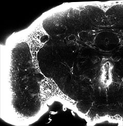
Serial confocal sections of the synaptic neuropile, visualized with repo-GAL4 and UAS-GFP
(32 MB, 871 x 894 pixels, 126 sections)


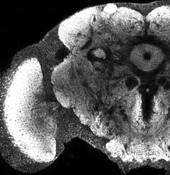
Serial confocal sections of the synaptic neuropile, visualized with the nc82 antibody
(36 MB, 871 x 894 pixels, 126 sections)

Side-by-side view of the neuropile and glial processes
(53 MB, 1256 x 894 pixels, 126 sections)


Montage view of the neuropile and glial processes
(43 MB, 871 x 894 pixels, 126 sections)